Flutter / AF

A 78 year old man is undergoing a second AF ablation procedure using an irrigated radiofrequency ablation catheter, under sedation only (no general anaesthesia or propofol). The transeptal puncture is performed without difficulty and catheters have been introduced into the left atrium. After delivering 22 minutes of RF energy the nurse reports a sudden drop in the systolic blood pressure, now 60/40. Which of the following actions is most appropriate?


  1. Rapidly administer 500mls of saline and continue the procedure
  2. Prepare the pericardiocentesis equipment while a transthoracic echo is performed.
  3. Call for a cardiac imaging specialist to perform a trans-oesphageal echo and advise on the next steps
  4. Administer an antidote to reverse the actions of the sedatives and analgesia given
  5. Stop the procedure and arrange an urgent contrast enhanced CT to rule out a retroperitoneal bleed

Answer : 2

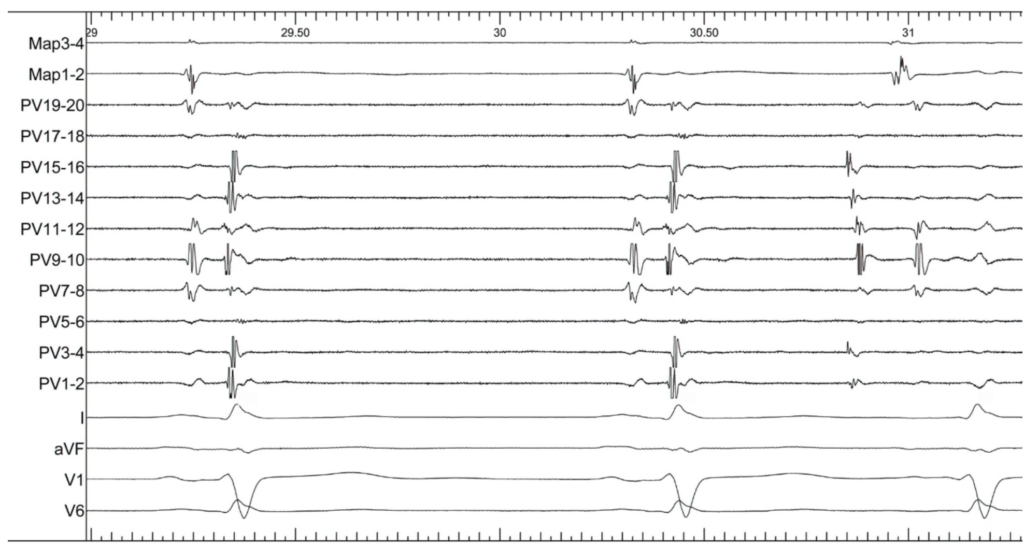
A 72 year old man with a history of paroxysmal AF and previous pumonary vein (PV) isolation procedure has a recurrence of symptoms and is brought back to the catheter lab for a repeat procedure. Mapping of the 4 PVs is performed. The tracing shows the the left superior PV. What conclusion can be drawn from this tracing?

Intra-cardiac electrograms are shown from a map catheter postioned in the left atrium (Map), and a duodecapolar catheter in the left superior pulmonary vein (PV). 4 ECG leads are shown.

  1. Ablation delivered via the map catheter at this site is likely to isolate the PV.
  2. There is entry but not exit block.
  3. There is exit but not entry block.
  4. There is neither entry nor exit block.
  5. The PV is isolated

Answer 4

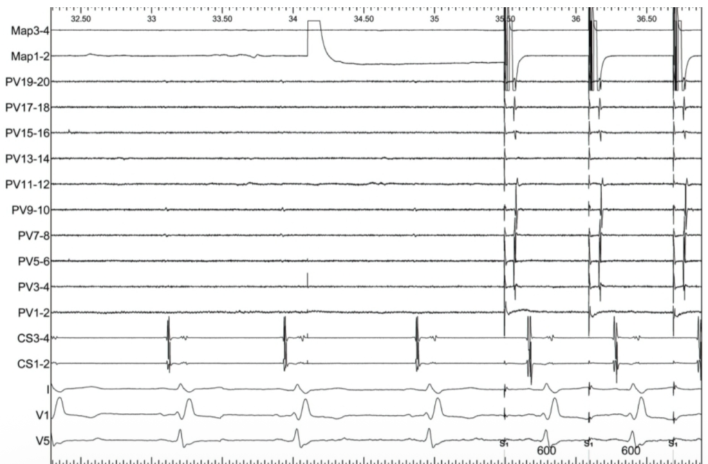
A 72 year old woman with diabetes but a structurally normal heart has paroxysmal AF. An ablation procedure is performed to isolate ipsilateral pairs of pulmonary veins using wide area circumferential RF ablation. While pacing from the ablation catheter the following tracing is recorded. Which statement is correct?


Intracardiac electrograms are shown from the ablation catheter (Map) positioned inside the right superior PV, a duodecapolar circular PV catheter in the right inferior PV and a quadripolar catheter in the coronary sinus with electrodes 3-4 positioned at the mouth of the CS. 3 ECG leads are shown. The paper sweep speed is 50mm/s.

1. Entry block in the right pulmonary veins is demonstrated

2. Further ablation is needed, targetting the earliest activation on the PV catheter

3. Dissociated PV potentials are seen, further ablation is not needed

4. The right superior and inferior PV are electrically isolated from each other

5. The right superior pulmonary vein is the likely source of the paroxysms of AF

Answer : 1

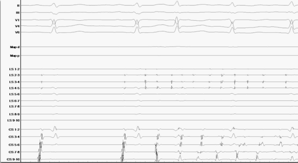
A 60 year old lady with recurrence of paroxysmal symptomatic paroxysmal AF. After previous PVI. What conclusion can be drawn from this tracing ?


1. Entry block of displayed PV is shown

2. Reconnection of PV

3. Reconnection of PV and initiation of AF

4. Exitblock of displayed PV is proven

5. Patient needs a pacemaker

Answer : 3

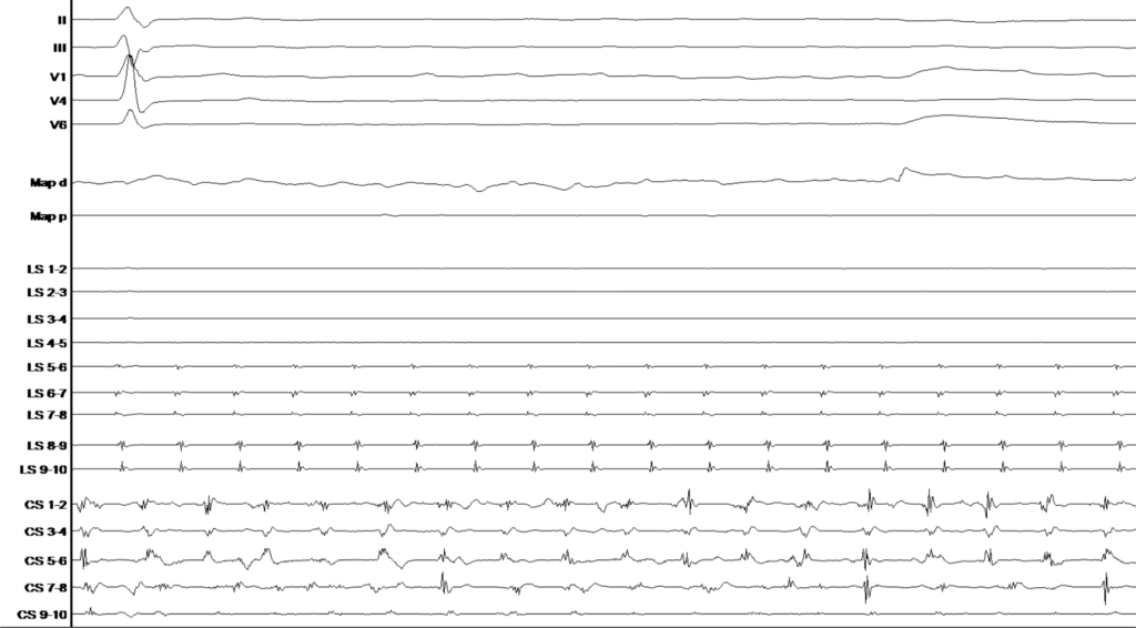
After the first PFA impulse of the LSPV the following tracing was obseverd.

What is most likely the causing of this observation and how could it be avoided ?


1. Accidental AVN ablation, put in a PM

2. Vagal reaction, stop procedure and use RF

3. Vagal reaction, administration of atropine before first impulse

4. Transient PM lead before ablation

5. AV-Block III°, PM Implantation

Answer : 3