SVT

Parahisian Pacing


Technique description:

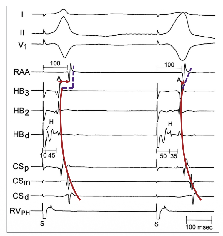
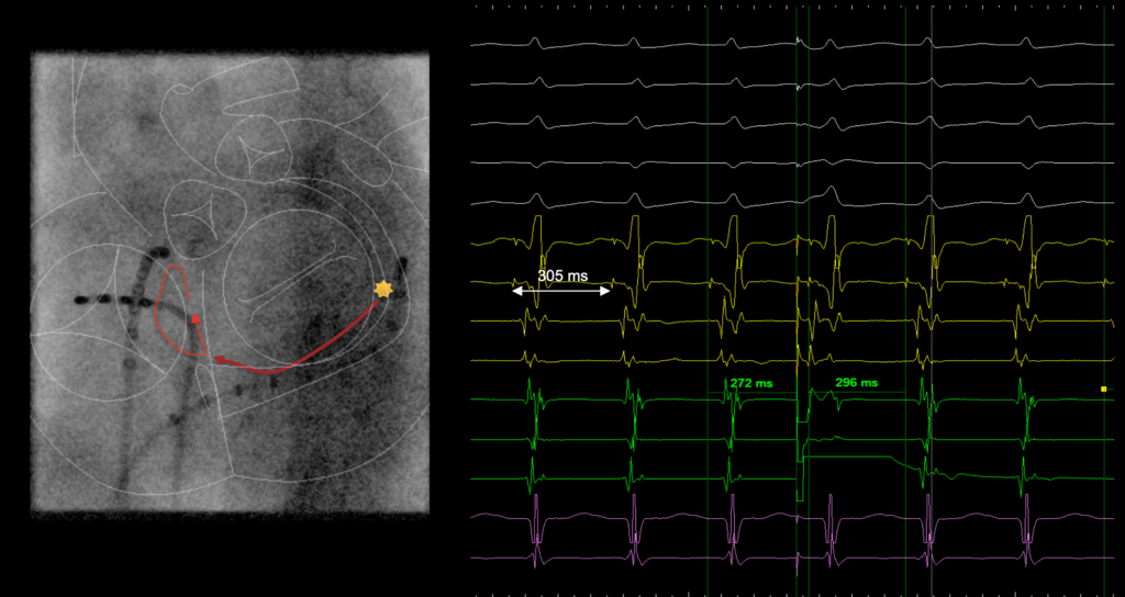
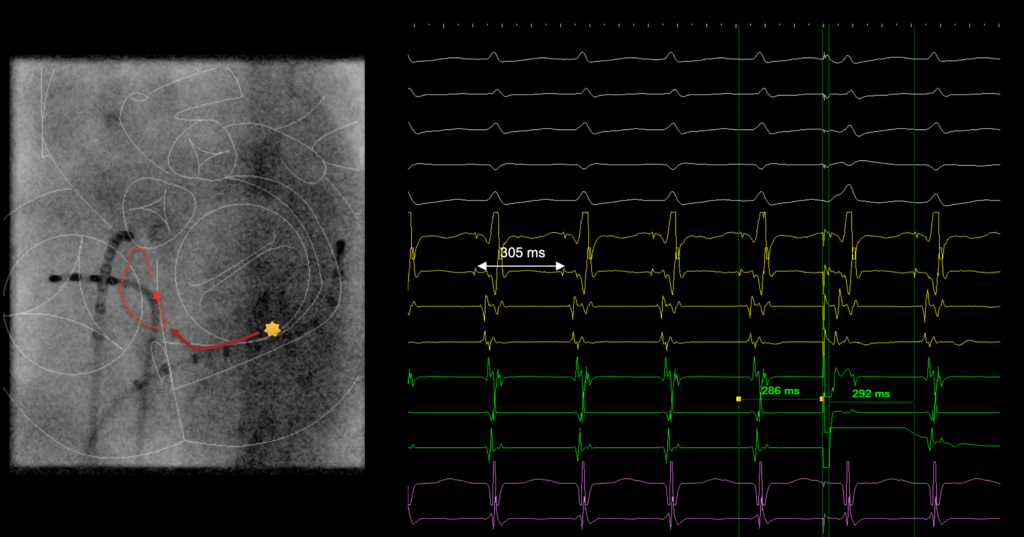
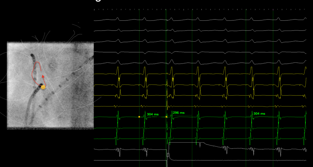
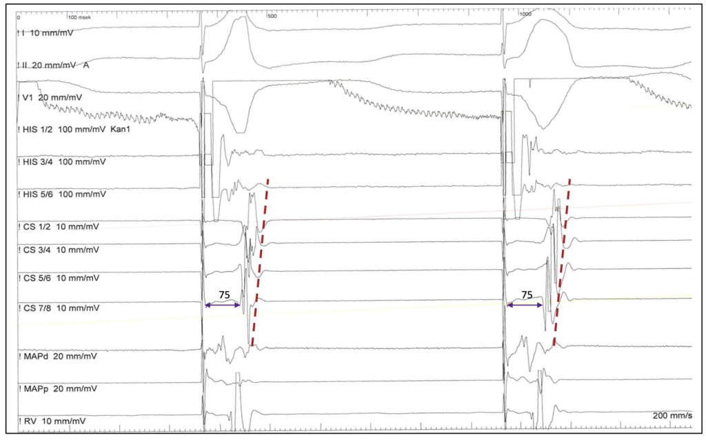
Ventricular pacing is performed adjacent to the His bundle / right bundle branch (HB-RB), initially at high output to capture both RV and HB-RB. The output is then decreased to lose HB-RB capture.


> An increase in S-A interval without change in His bundle-atrial interval or atrial activation sequence indicated exclusive retrograde AVN conduction


> Loss of HB-RB capture without change in stimulus -atrial (S-A) interval or atrial activation sequence indicated exclusive retrograde AP conduction.


> A change in atrial activation sequence indicated the presence of both retrograde AP and AVN conduction.

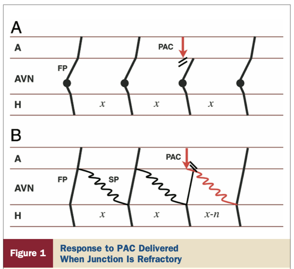
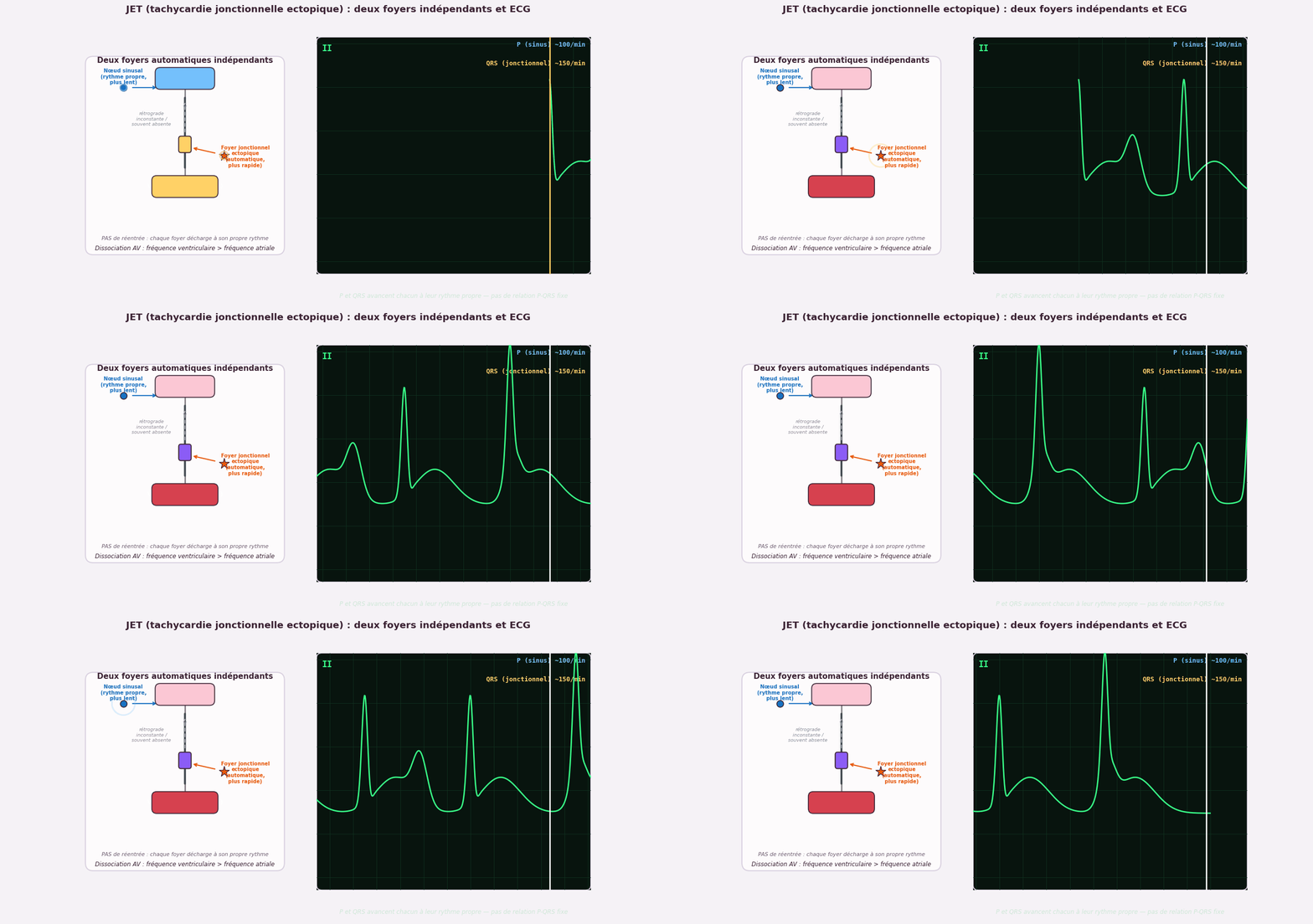
PAC during the SVT -> AVRNT VS JET

Response to premature atrial complex (PAC) delivered when junction is refractory (local atrial activation from PAC occurs at or after His activation). (A) Response in junctional tachycardia: a PAC delivered at a time the junction focus has already depolarized blocks at the atrioventricular node (AVN) and is unable to influence the immediate or the next junction beat. Solid circles represent junction focus. Black lines show conduction through AVN, His (H), and atrium (A). (B) Response in atrioventricular node re-entry tachycardia (AVNRT): a similarly timed PAC can influence the next beat of AVNRT by early engagement of the slow pathway. Black lines show conduction through AVN, His (H), and atrium (A), and red lines show PAC and its response. Although this figure shows advancement of the next beat (x-n), delay of the next beat or termination of tachycardia are also specific to AVNRT. Red arrow indicates PAC. FP  fast atrioventricular node pathway; SP  slow atrioventricular node pathway; x and x-n  H-H intervals.

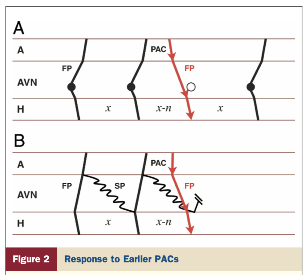
(A) Response in junctional tachycardia (JT): the open circle represents the anticipated JT beat timing if no PAC were delivered. An early PAC advances the immediate JT beat and His timing by atrioventricular (AV) nodal fast pathway activation and JT continues. (B) Response in AVNRT: an early PAC may advance the immediate His by activation of the AV nodal fast pathway. However, that makes the fast pathway refractory and unavailable for retrograde conduction, terminating the AVNRT circuit. Red arrow indicates PAC and its response. Legends and abbreviations as in Figure 1.

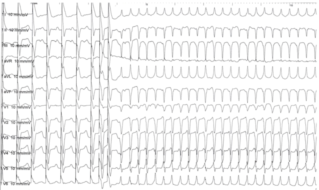
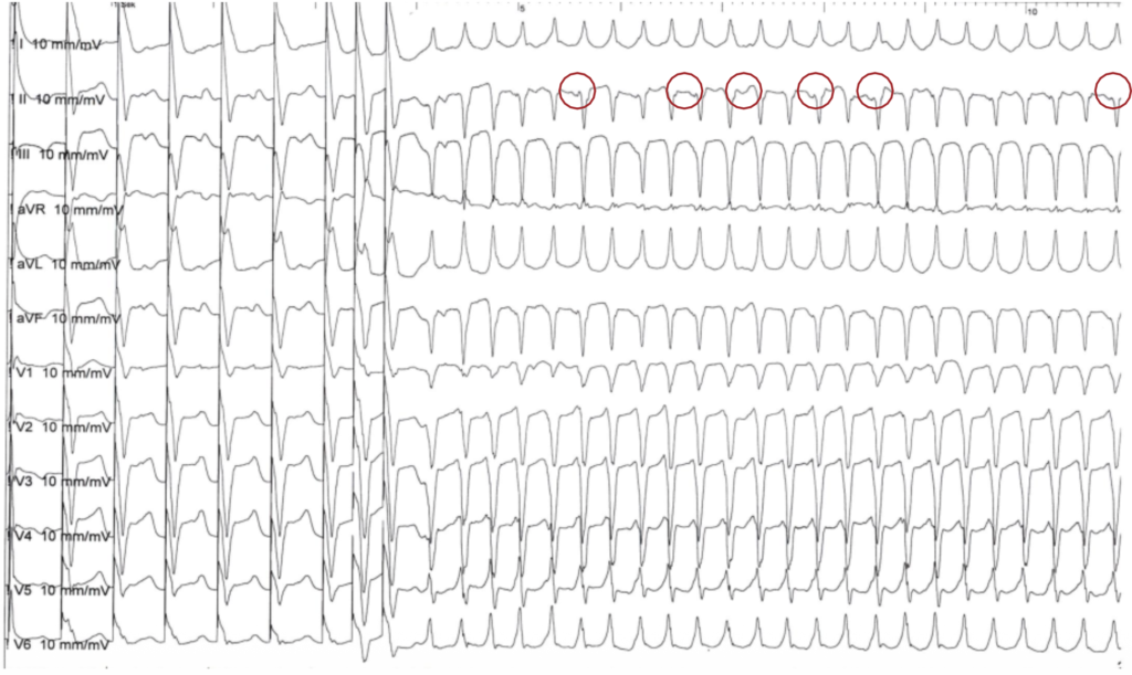
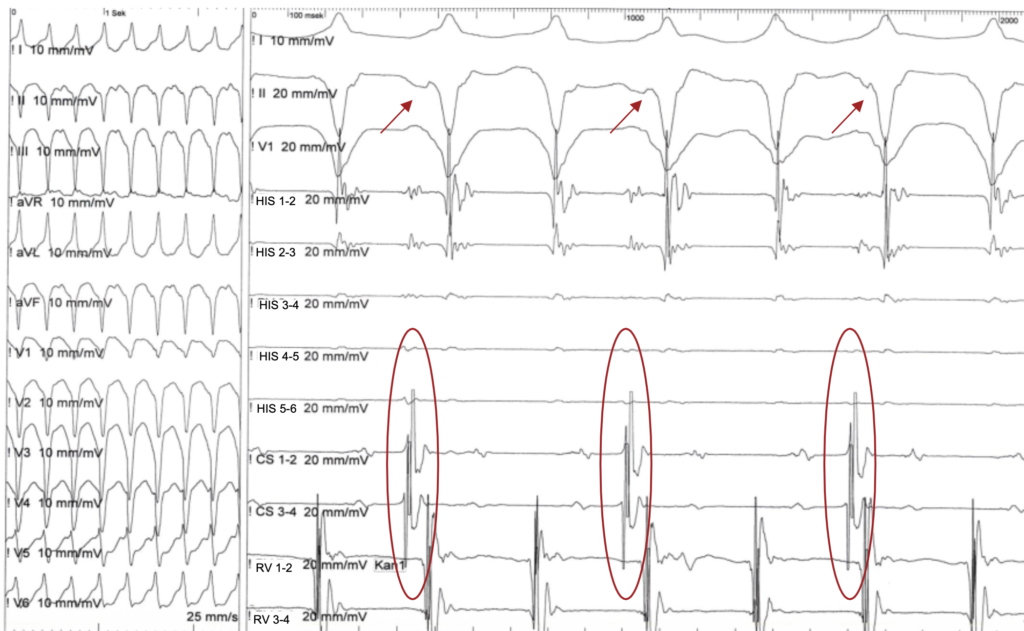
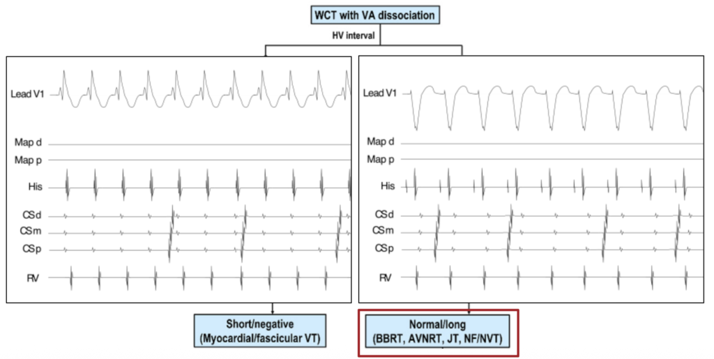
2. Wide complex tachycardia with VA dissociation

What is your diagnosis ?

1. SVT with aberrancy
2. Preexcited AT
3. Antidromic AVRT
4. Ventricular tachycardia
5. BBRVT/ILVT
6. I don´t know show me the endocardial signals

What is the next step ?

-> His recording

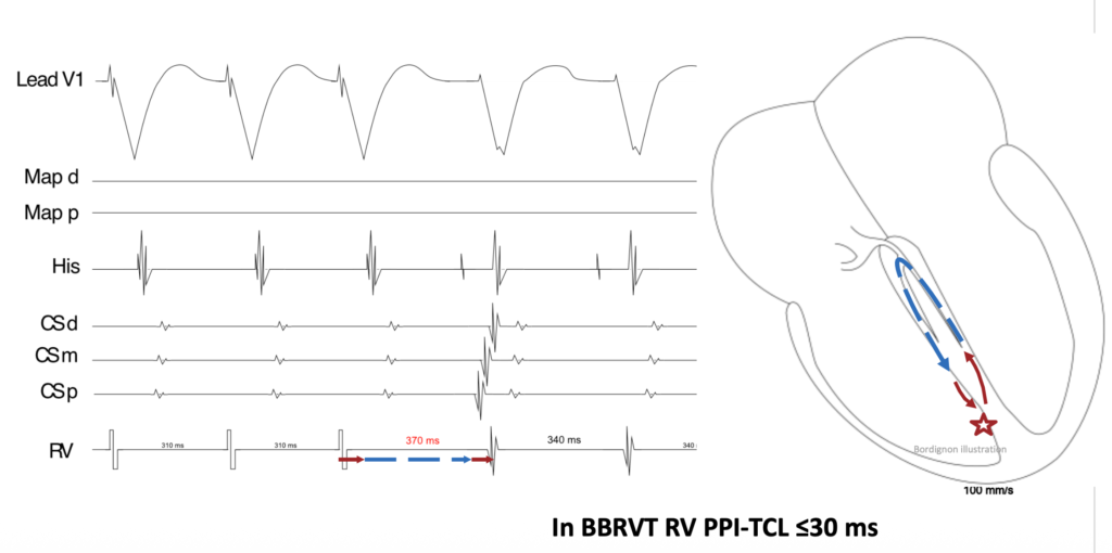
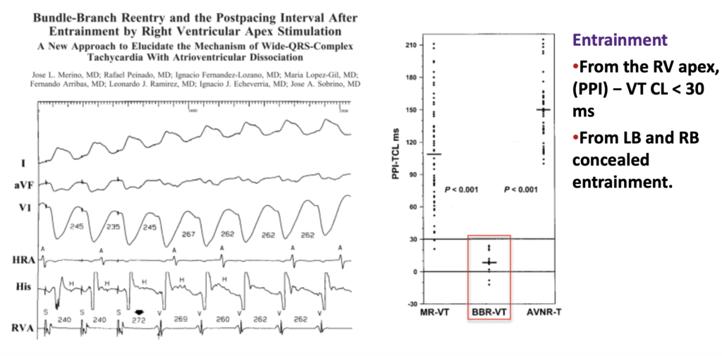
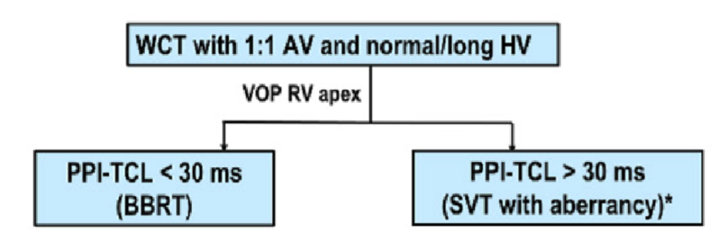
CRITERIA TO DIAGNOSE A BBRVT : 

EP Criteria #1: HIS

  • A His potential precedes the V
  • HV interval during SR is usually (!) prolonged

EP Criteria #2: HH-VV

  • HH Variations precede VV Variation

EP Criteria #3: HV

  • HV interval during BBR-VT with a LBBB pattern is usually similar to or longer than that during sinus rhythm

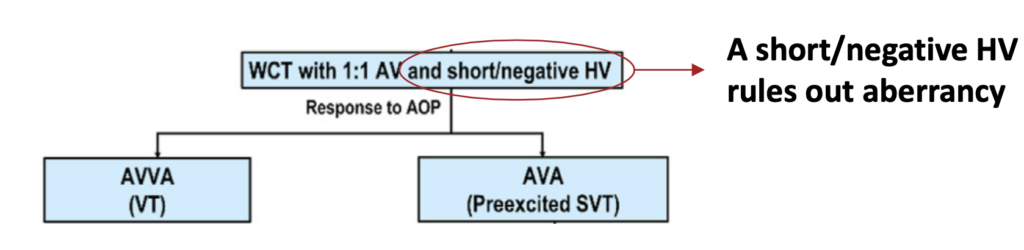
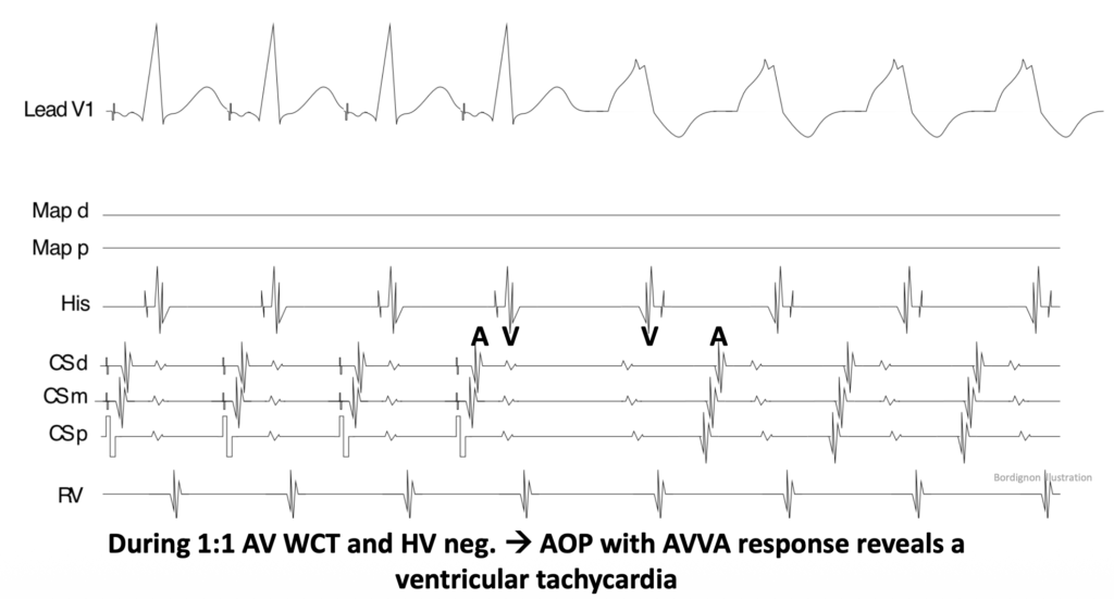
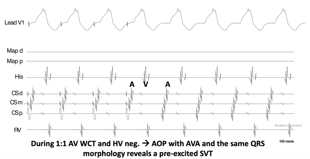
3. Wide complex tachycardia 1:1 AV

4. Study Cases

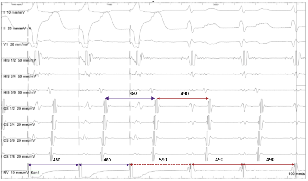
What is the diagnosis ?

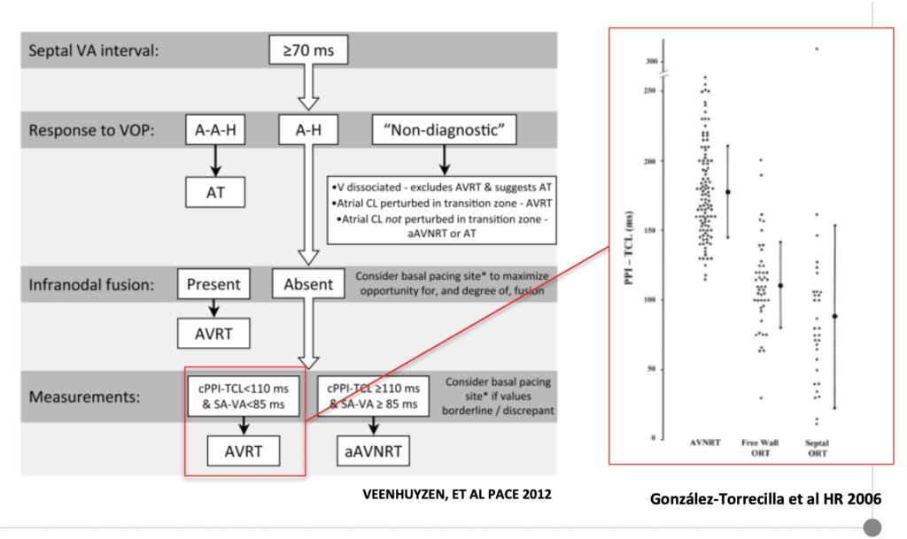
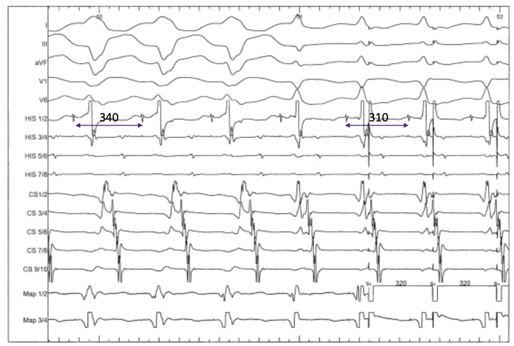
-> RA > 70 ms : AVRT, AT, aAVNRT

-> CS1,2 first last CS9,10 : AVRT

-> Increase CL with LBB > ipsilateral AP

-> H before V > orthodromic AP


Conclusion : ORT via left AP  

-> Loss of HB-RB capture without change in stimulus -atrial (S-A) interval or atrial activation sequence indicated exclusive retrograde AP conduction


Conduction via an accessory pathway only

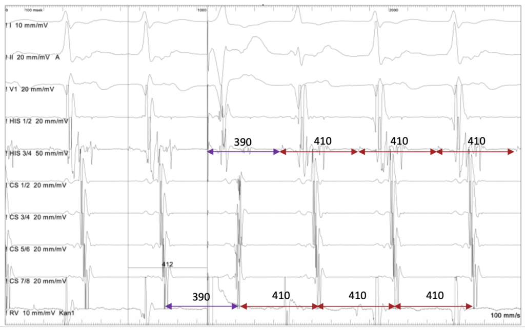
What is the diagnosis ?

  • VAAV response
  • First activation CS1,2

-> Left sided AT

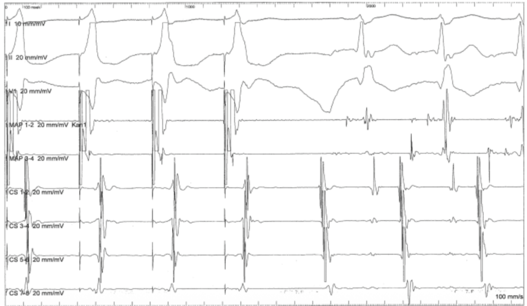
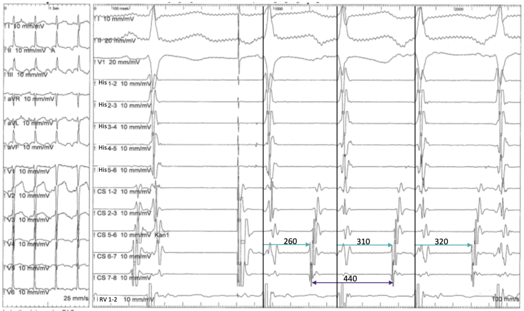
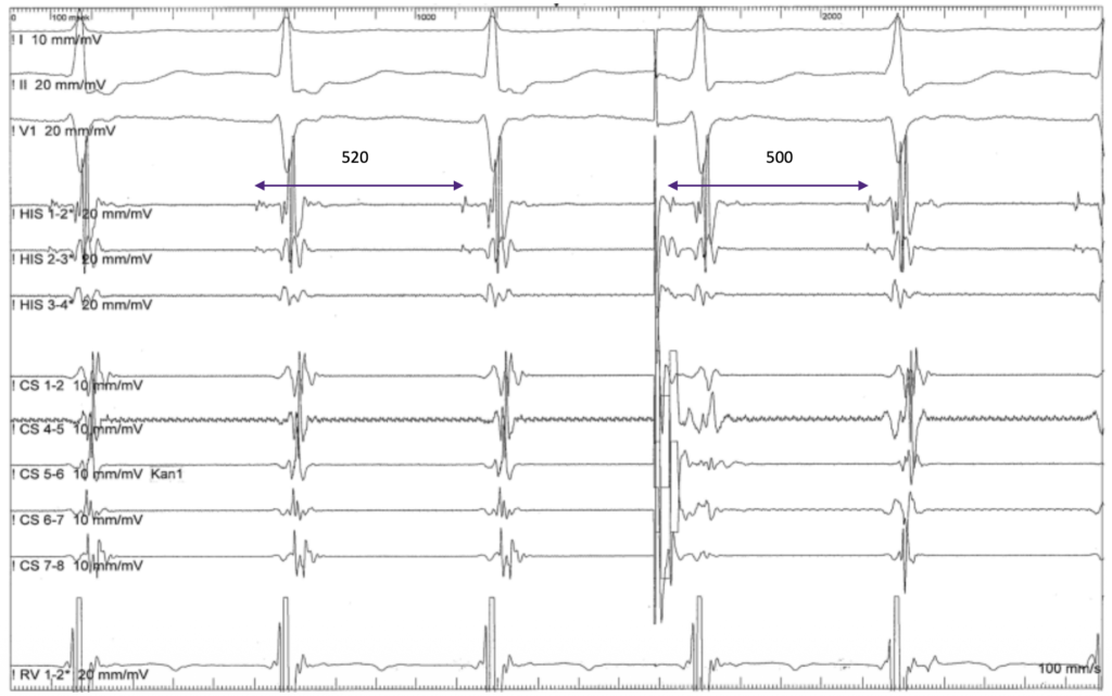
What is the diagnosis ?

  • 1:1 tachycardia
  • Visible His
  • PVC during His refractory (ECG fusion) > A advanced
  • Changement of atrial activation with PVC

-> ORT with two accessory pathways, one as bystander

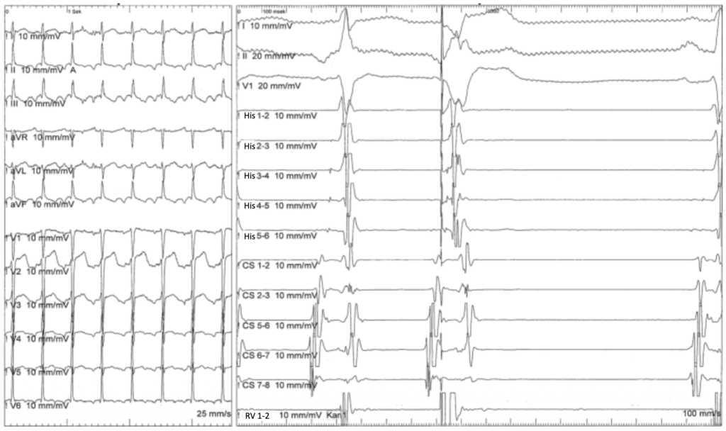
  1. Slow slow AVNRT
  2. Fast-slow AVNRT
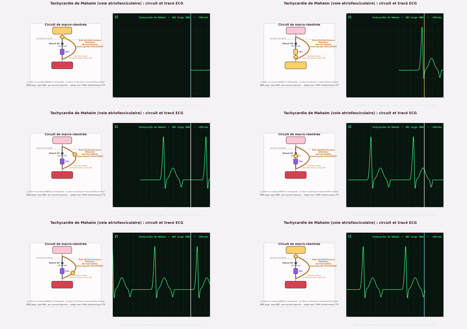
  3. ORT via a Mahaim fiber
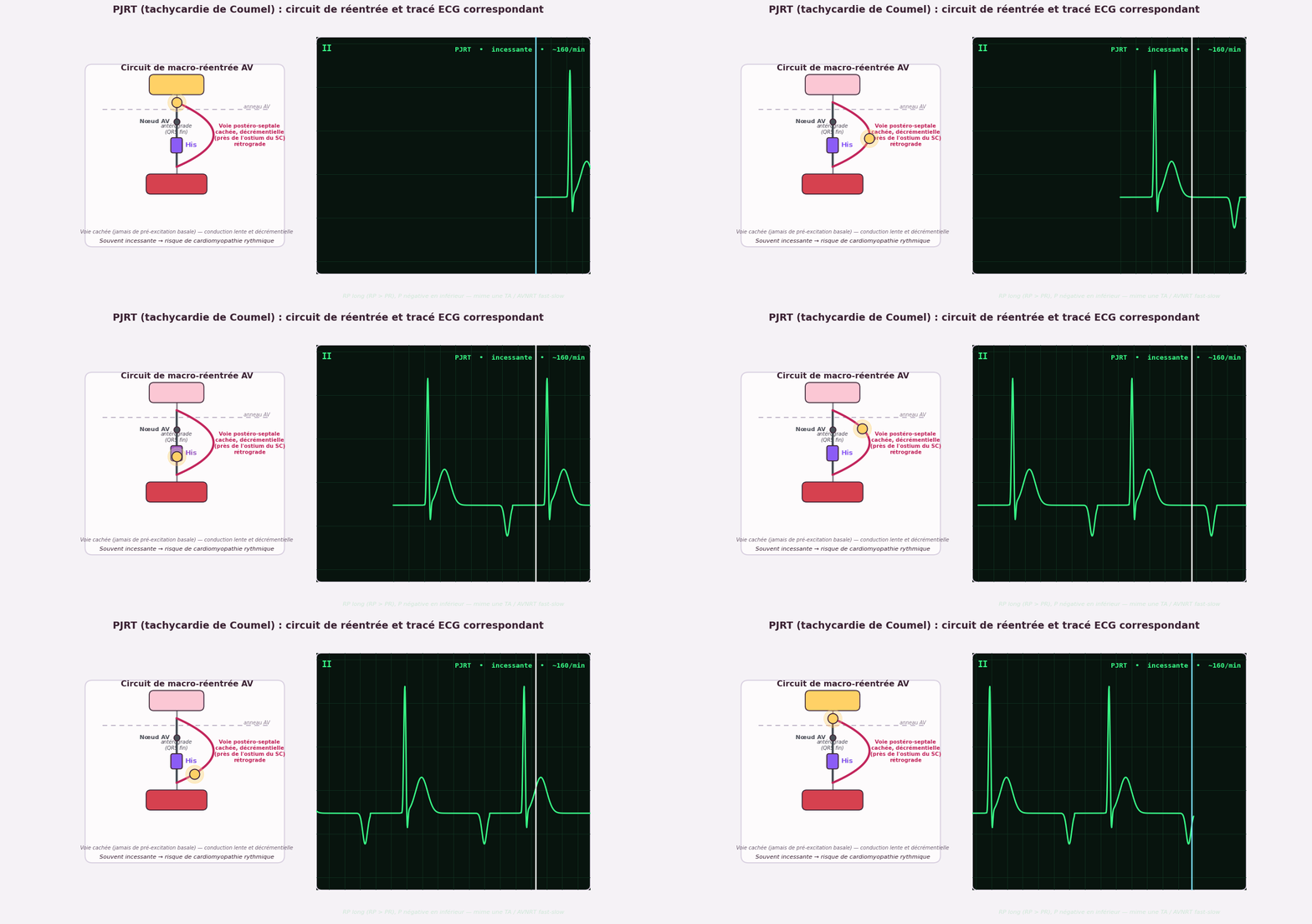
  4. PJRT
  5. AT

VS

A His-refractory PVC that terminates the tachycardia points to an accessory pathway with retrograde-only conduction. This excludes an antegradely conducting pathway — i.e., it rules out a Mahaim fiber. What remains is PJRT, which conducts in a retrograde, decremental (slow) fashion

1. Typical AVNRT

2. Atypical AVNRT

3. ORT

4. AT 1:1

5. Atrial Flutter

> ORT

1. Typical AVNRT

2. JET

3. ORT

4. AT

5. Atrial Flutter

VS

The PAC arrives during the His refractory period. If this were JET, the V would not be advanced. Here the V is advanced because conduction proceeds down the slow pathway — this confirms AVNRT.
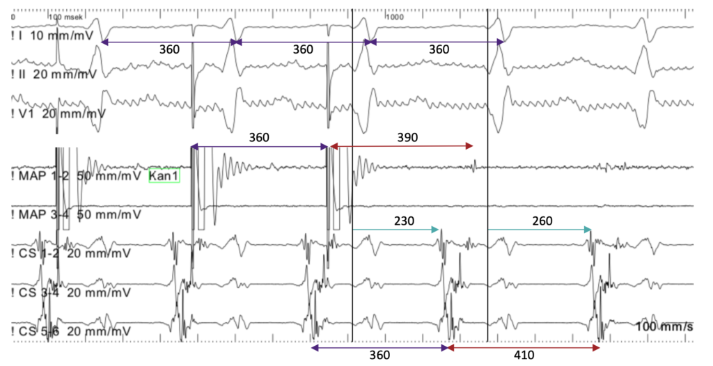
We are pacing from the left atrium

What is the diagnosis?

1. Typ. AVNRT

2. Atyp. AVNRT the SP is involved

3. AVRT: the CS Activation is distal to proximal

4. AT 1:1, no VA Linking

5. ORT via a decremental accessory pathay

-> left sided AT good spot to ablate PPI-TCL negative