Long Qt syndrome

1. Physiology

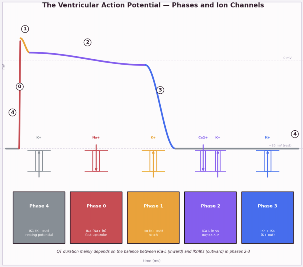
Picture the heart cell as a nightclub. At rest (phase 4), it’s calm and quiet: the music’s off, and only a few bouncers (the IK1 channels) let a trickle of K+ out to keep things settled at -85 mV. Then the electrical signal arrives and it’s chaos: every Na+ door bursts open at once (phase 0), a crowd of Na+ rushes in, and the voltage rockets up in a fraction of a millisecond — that’s the sharp vertical spike. Right after, a brief lull (phase 1): a few K+ slip back out, like a bouncer easing the crowd a bit, which gives that little notch on the curve.

Then comes the plateau (phase 2), the longest part of the night: a real tug-of-war between Ca2+ still trickling in (through ICa-L — and this same calcium is what makes the muscle actually contract) and K+ quietly starting to leave (via IKr, IKs). As long as these two flows roughly balance out, the party stays steady on that plateau. Eventually K+ wins out (phase 3): everyone heads for the exit, the room empties, and the voltage drops back to -85 mV — that’s repolarization. And then it’s back to phase 4, ready for the next beat.

Same nightclub, but now a few of the doors are faulty.

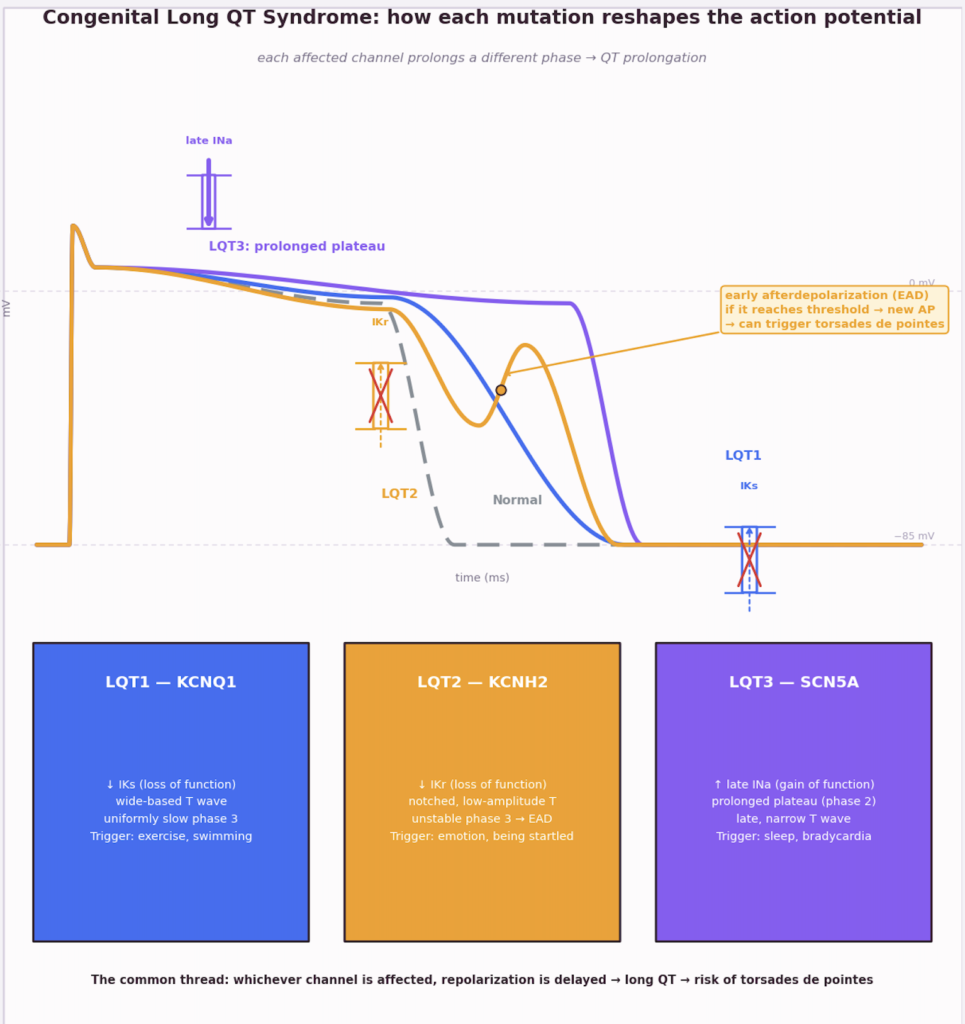
LQT1: the IKs exit door is half-jammed. People can’t leave as fast as they should late in the night, so phase 3 stretches out — but evenly, just a slow, uniform closing. Since exercise is exactly when you need that exit to speed up, this one tends to decompensate with sport or swimming.

LQT2: the IKr door is outright glitchy, opening and sticking at random. The exit turns chaotic, and sometimes the door pops back open and lets a second small wave of people back in before the room has fully cleared — that’s exactly the bump you see on the orange curve (the EAD, early afterdepolarization). If that second wave is strong enough, it can fire off a stray new action potential, which is the classic starting point for torsades de pointes. Typical trigger: a sudden noise, an alarm going off, a strong emotion.

LQT3: here the problem isn’t the exit but an entry door (Na+) that won’t shut properly. A steady trickle of Na+ keeps leaking in throughout the plateau, so the room stays “lit up” longer — hence the abnormally prolonged plateau. That leak shows up mostly when everything’s quiet, so sleep and bradycardia are the classic risk moments.

The common thread across all three: the room always takes longer to close down (repolarize), so the QT stretches out on the ECG — and the more unstable and unpredictable that closing gets (as in LQT2), the higher the risk of torsades.

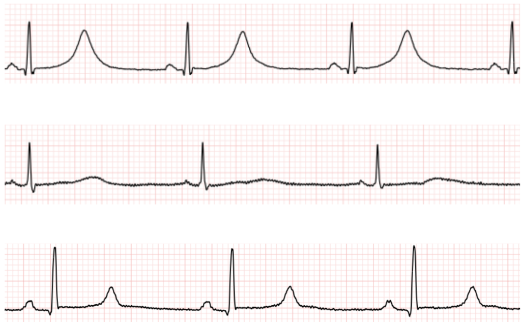
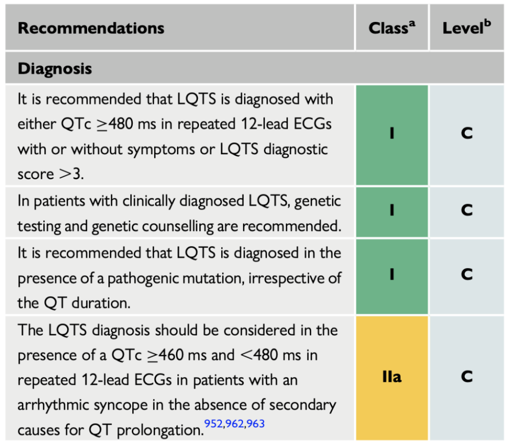
2. Diagnosis

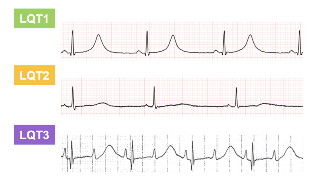
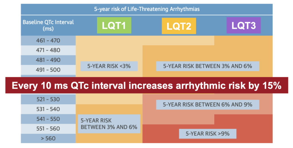
Long Qt syndrome types

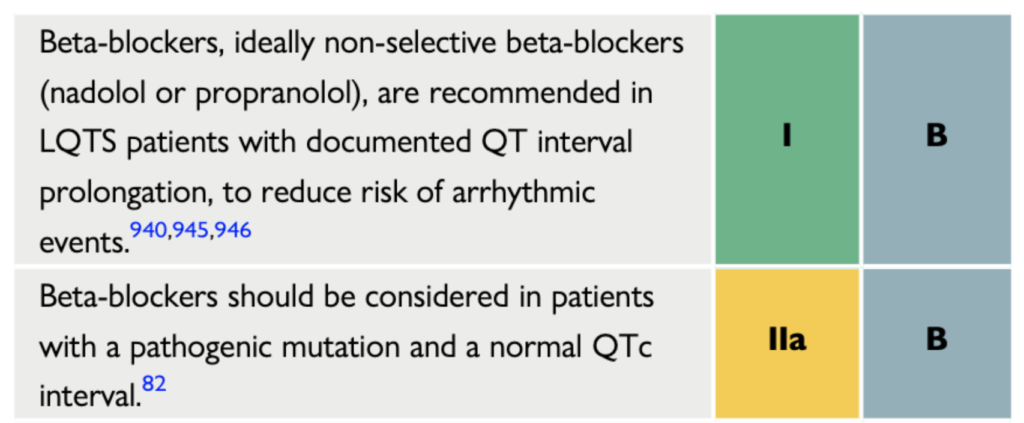
Nadolol dosage 1 mg/kg/day
Propranolol 1 mg/kg TID
Mexiletine dose 8mg/kg/day

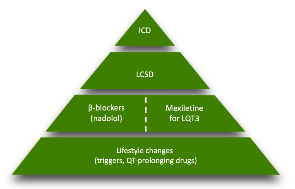
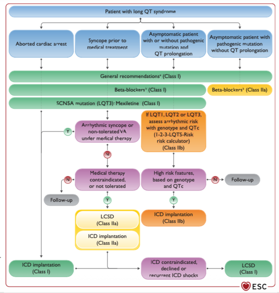
When to implant an ICD ?

Study cases

A 27‑year‑old woman with congenital long‑QT syndrome has a pathogenic SCN5A variant (LQT3). Her QTc is markedly prolonged despite maximally tolerated propranolol. Which therapy is specifically indicated for patients with LQT3 and a prolonged QT interval?


A. Increase the β‑blocker dose further


B. Start mexiletine


C. Add a calcium channel blocker


D. Initiate potassium supplementation


E. Start flecainide for atrial arrhythmia suppression

-> Start Mexiletine

A 35‑year‑old man is newly diagnosed with congenital LQTS (QTc 490 ms). He is asymptomatic, and genetic testing is pending. He asks about further tests and indications for an implantable cardioverter‑defibrillator (ICD). Which statement best reflects ESC guideline recommendations?


A. Routine epinephrine challenge is recommended to guide therapy


B. Risk of arrhythmia should be estimated based on genotype and QTc duration before therapy


C. Selective β‑blockers (e.g., metoprolol) are preferred over propranolol or nadolol


D. All asymptomatic LQTS patients should receive an ICD


E. Mexiletine is first‑line therapy regardless of genotype

-> calculate arrhythmic risk in LQTS based on genotype and QTc duration before initiating therapy. Routine epinephrine challenge is not recommended. Non‑selective β‑blockers are the recommended first‑line agents. ICD implantation may be considered in asymptomatic patients only when they have a high‑risk profile despite genotype‑specific therapy

A 27 yo patient presents after experiencing syncope to our outpatient clinic. Genetic testing shows a pathogenic SCN5A mutation. What is the best strategy?


A) Likely reflex syncope. Follow-up


B) ICD implantation.


C) Initiate betablockers and consider mexiletine.


D) Implantable loop recorder.


E) Cardioneuroablation

C