Synapse the ECG with Dr. Synapse
ECG – Green JM, Chiaramida AJ. ECG – 12 – Lead EKG Confidence: A Step by Step Guide. 1st ed. New York: Springer

Atrial and ventricular pacing.
The pacing spike hits the T wave, causing V Tach, and V fib.
ECG – Green JM, Chiaramida AJ. ECG – 12 – Lead EKG Confidence: A Step by Step Guide. 1st ed. New York: Springer

Atrial tachycardia (AT) or supraventricular tachycardia (SVT) is a regular rhythm at a rate of 160 to 260. A P wave, if present (P′), appears differently from the normal P wave (P).
The heart rate increases from 91 to more than 160 bpm.
Atrial tachycardia is usually associated with increased sympathetic activity. It can be seen in normal and abnormal hearts.
ECG – Green JM, Chiaramida AJ. ECG – 12 – Lead EKG Confidence: A Step by Step Guide. 1st ed. New York: Springer

Third-degree AV block
ECG – Green JM, Chiaramida AJ. ECG – 12 – Lead EKG Confidence: A Step by Step Guide. 1st ed. New York: Springer

Torsade de pointe
ECG – Green JM, Chiaramida AJ. ECG – 12 – Lead EKG Confidence: A Step by Step Guide. 1st ed. New York: Springer

2nd degree AV block (2° AVB) Type I, Wenckebach, with 4 to 3 conduction.
The PR interval gradually increases until a QRS is dropped. There are 4 P waves to 3 QRS complexes, thus a 4 to 3 ratio
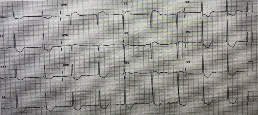
ECG – Dr. Kamel Kamel (General Practitioner):
70-year-old patient with chest pain and hypertension.

Regular sinus rhythm
- QRS > 120 ms: Here, 150 ms
- Due to delayed depolarization of the right ventricle:
-> RSR’ pattern in leads V1-V3, resembling an “M” shape
-> Wide S wave in lateral leads DI, aVL, V5-6
Conclusion: This is a complete right bundle branch block (RBBB).
Regarding the chest pain -> it will be necessary to clarify the onset and type of chest pain.
ECG – Green JM, Chiaramida AJ. ECG – 12 – Lead EKG Confidence: A Step by Step Guide. 1st ed. New York: Springer

PVC, followed by 3:2 and 2:1 Wenckebach.
ECG – Green JM, Chiaramida AJ. ECG – 12 – Lead EKG Confidence: A Step by Step Guide. 1st ed. New York: Springer

Artifact
This ECG demonstrates a rhythm recorded on a two-channel recorder. Both leads are recorded at the exact same time, so any event on the top strip occurs at exactly the same time as an event exactly below it.
The rhythm on top appears to be ventricular tachycardia until compared with the bottom strip, which shows sinus rhythm. Manipulation of electrodes can cause this.
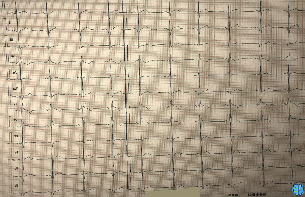
ECG – Dr Synapse :
30-year-old patient admitted to the emergency department for syncope.

Appearance of a right bundle branch block, best seen in leads V1 and V2.
- The epsilon wave is clearly visible in V1, just after the QRS complex.
- Negative T waves in leads V1-V2-V3.
This is likely a case of ARVD: Arrhythmogenic Right Ventricular Dysplasia.
ECG – Green JM, Chiaramida AJ. ECG – 12 – Lead EKG Confidence: A Step by Step Guide. 1st ed. New York: Springer

Atrial flutter with 6:1 conduction.
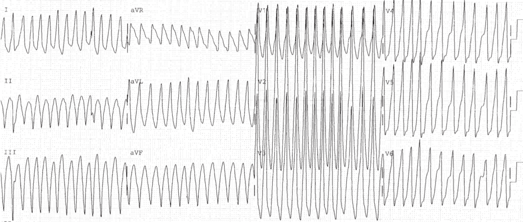
ECG – Green JM, Chiaramida AJ. ECG – 12 – Lead EKG Confidence: A Step by Step Guide. 1st ed. New York: Springer

Torsade de Pointe (polymophous ventricular tachycardia) is a form of ventricular tachycardia. The diagnostic hallmark of torsade is a change in direction of the complexes.
The episode of ventricular tachycardia begins with the first three QRS complexes pointing downward, and the next three pointing upward. This is associated with long QTc interval. Drugs, hypokalemia, and hypocalcemia are common causes.
ECG – Green JM, Chiaramida AJ. ECG – 12 – Lead EKG Confidence: A Step by Step Guide. 1st ed. New York: Springer

Atrial flutter is a regular atrial rhythm characterized by atrial flutter waves. Lead II frequently demonstrates the classic sawtooth appearance of the flutter waves, some of which are hidden in the ST segment.
Atrial flutter usually is a regular rhythm at a rate of 240 to 340 with flutter waves, but not every flutter wave produces a QRS. The number of flutter waves to QRS complex is described as flutter with varying 3 to 1 and 2 to 1 conduction.
The atrial rate (230) and ventricular rate (88 to 115) should be described separately. Atrial flutter frequently is associated with underlying heart or lung disease.
ECG – Dr Synapse

What stands out on this ECG is:
- Bradycardia without P waves: This is a junctional rhythm at 50 bpm -> consistent with sinoatrial block.
- ST segment with a peculiar “scooped” shape: This is typical of digoxin effect/toxicity.
Conclusion: The patient likely has digoxin toxicity.
ECG – Green JM, Chiaramida AJ. ECG – 12 – Lead EKG Confidence: A Step by Step Guide. 1st ed. New York: Springer

1°AV block, 2:1 2°AVB.
ECG – Green JM, Chiaramida AJ. ECG – 12 – Lead EKG Confidence: A Step by Step Guide. 1st ed. New York: Springer

Idioventricular rhythm is a slow ventricular escape rhythm characterized by wide QRS complexes, typically in a regular slow pattern with no underlying pacing in the ventricles.
This is associated with underlying heart disease and can be worsened by beta blockers or calcium channel blockers.
ECG – Green JM, Chiaramida AJ. ECG – 12 – Lead EKG Confidence: A Step by Step Guide. 1st ed. New York: Springer

2 dropped P waves.
2°AV block, Type II.
ECG – Green JM, Chiaramida AJ. ECG – 12 – Lead EKG Confidence: A Step by Step Guide. 1st ed. New York: Springer

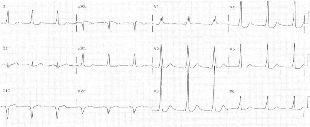
Short PR interval + Delta Wave = Wolff Parkinson White
ECG – Green JM, Chiaramida AJ. ECG – 12 – Lead EKG Confidence: A Step by Step Guide. 1st ed. New York: Springer
