LV lead placement

1. Coronary Sinus anatomy

The first thing to visualize carefully is the anatomy of the coronary sinus in RAO 30° and LAO 30° (ideally 40°).
This is, in my view, the most important step: which coronary sinus branch are you going to target?
In the RAO view, the atrium appears on the left side of the screen and the ventricle on the right.
This projection helps determine whether a structure is basal, mid, or apical.
The coronary sinus ostium is located posterior to the tricuspid valve annulus.
From this posterior–basal region, the coronary venous system branches as follows:
  • Middle cardiac vein – runs along the posterior interventricular groove.
  • Posterolateral vein – courses toward the posterolateral left ventricular wall. (PRIMARY TARGET for LV pacing in CRT.)
  • Lateral (marginal) vein – follows the lateral left ventricular wall. (KEY target for LV pacing.)
  • Anterolateral vein – projects toward the anterolateral wall.
  • Great cardiac vein – ascends in the anterior interventricular groove and then continues toward the coronary sinus.
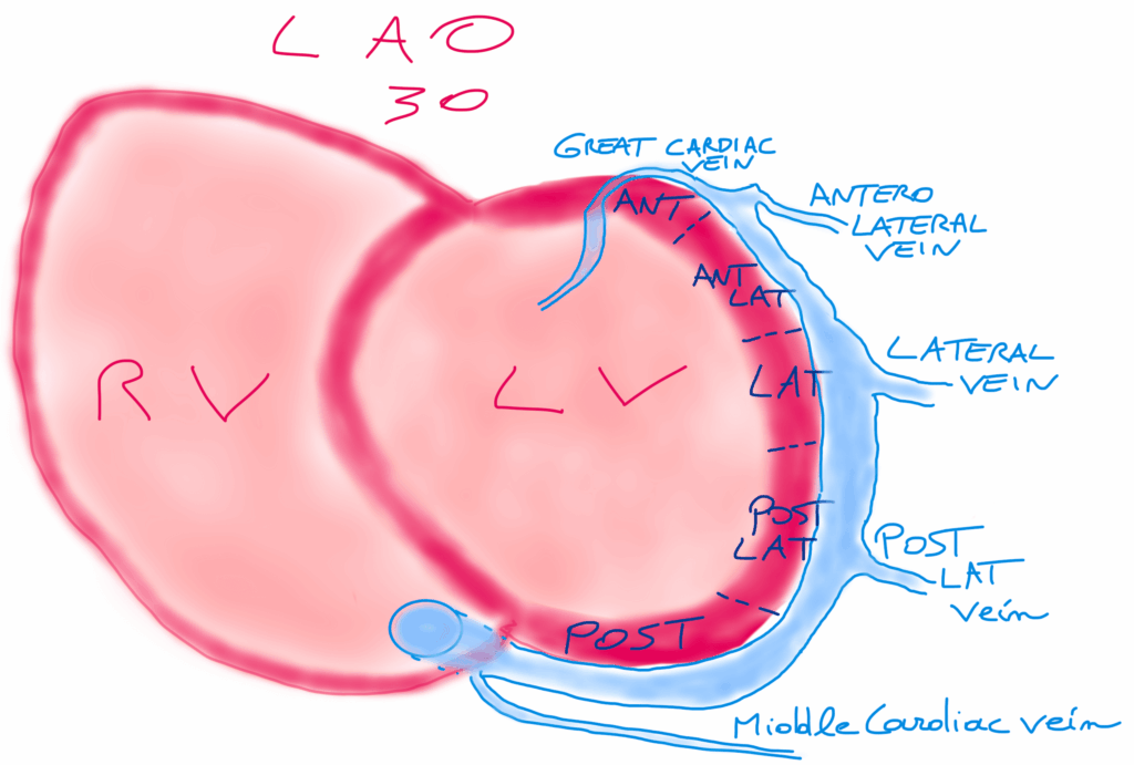
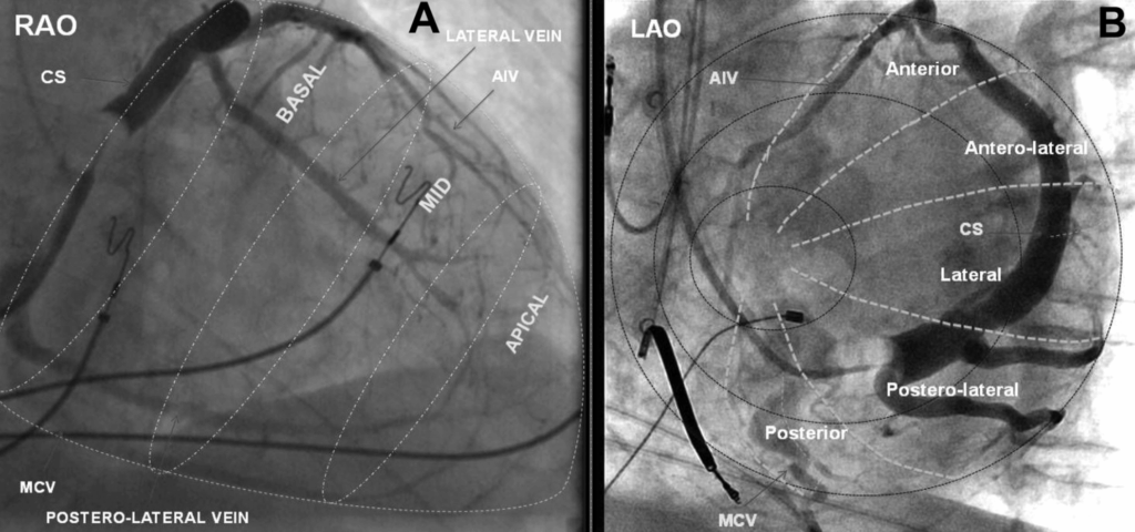
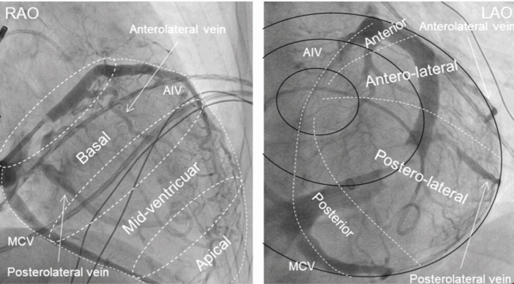
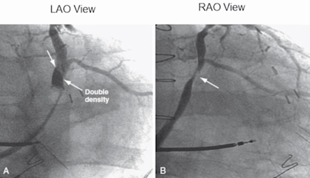
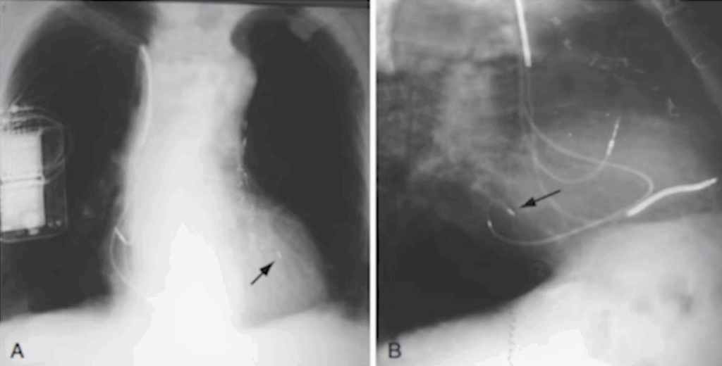
In the LAO view, you visualize the two “discs” of the right ventricle, with the right-sided structures on the left of the screen and the left-sided structures on the right.
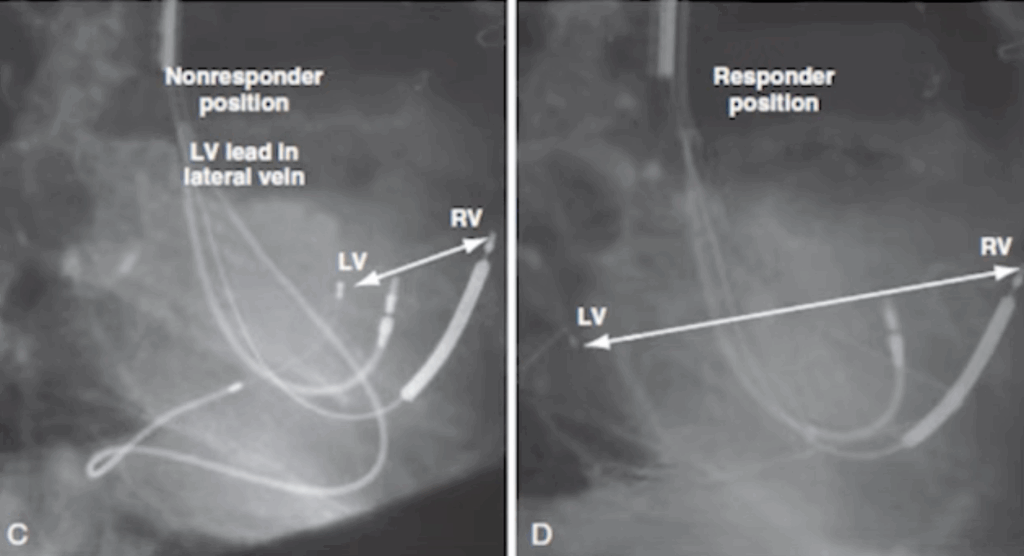
For LV pacing, the goal is to place the LV lead as far as possible from the RV lead to achieve optimal ventricular synchrony.
This is also the reason why the RV lead is placed first: once its position is fixed, you can determine exactly where the LV lead should be positioned to maximize separation and improve resynchronization.
The LAO view is essential, because it allows you to assess whether your chosen LV pacing site is likely to deliver good long-term CRT response.

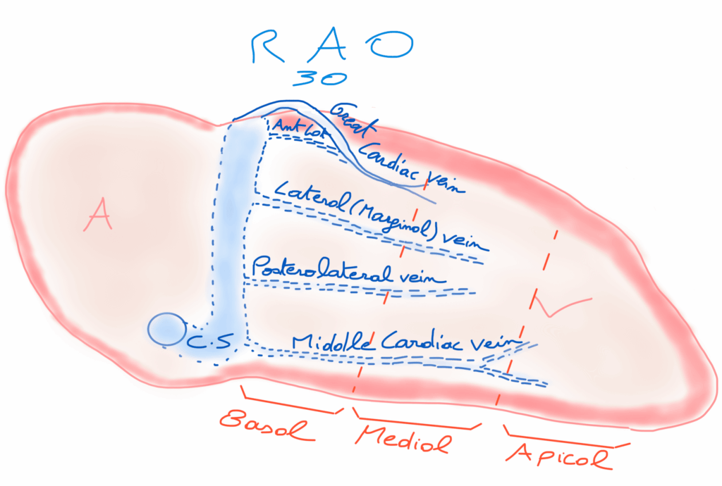
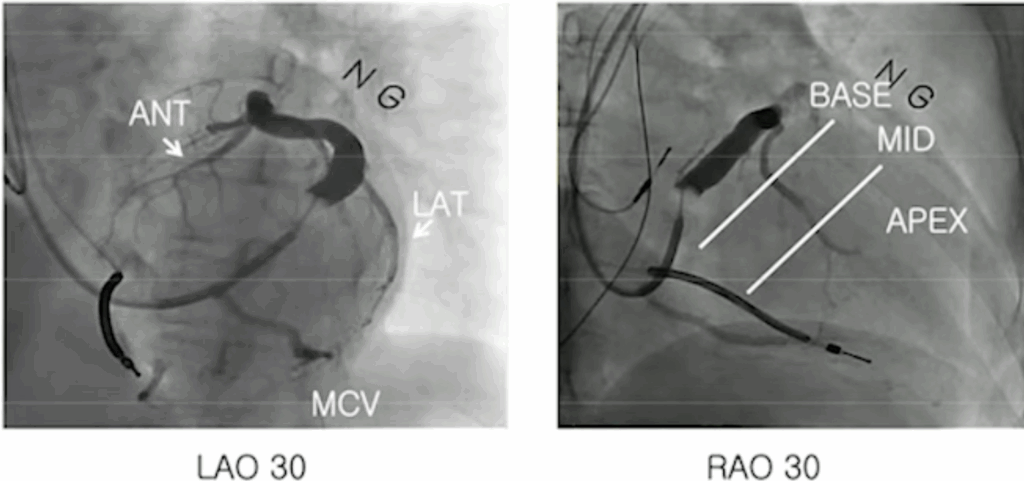
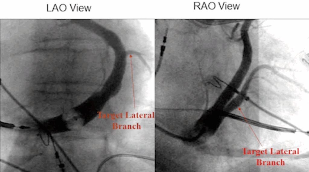
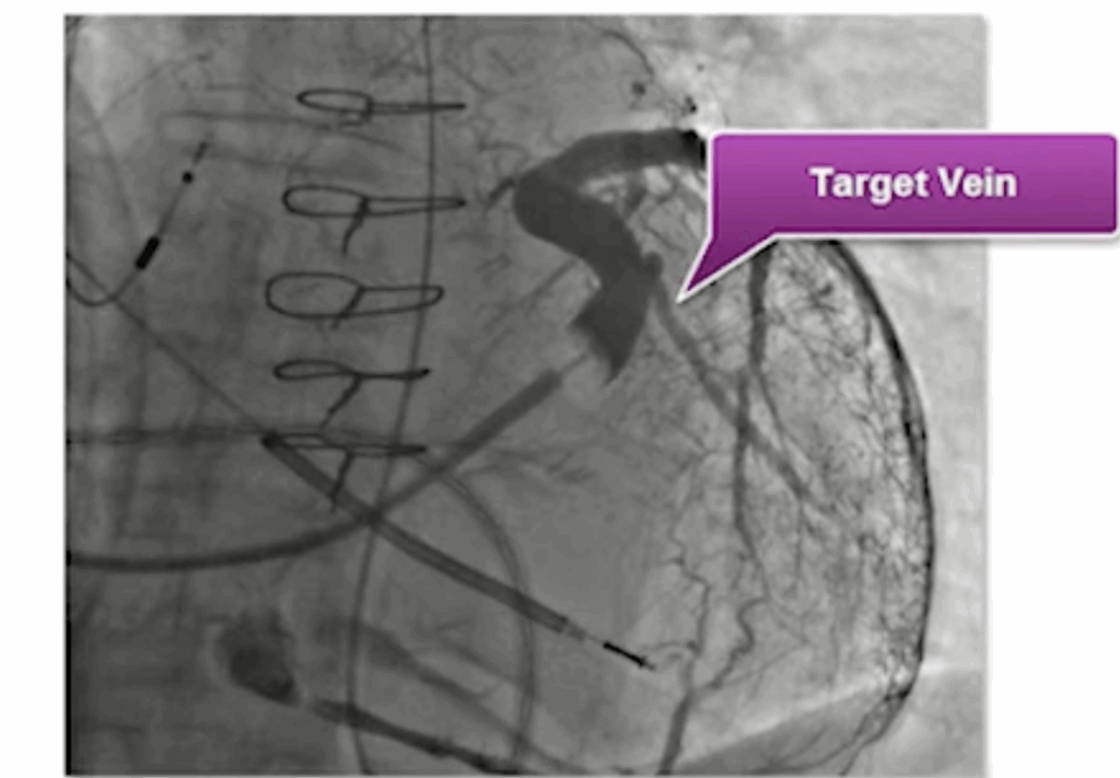
Practically, you target lateral venous branches located between 2 and 5 o’clock, ideally the posterolateral or lateral veins, which usually provide the most favorable electrical and mechanical delay.

2 - Target the best LV lead position

After positioning the RV lead first, the next step is to cannulate the coronary sinus and inject contrast in both LAO and RAO views to identify the optimal target vein for placing the LV lead.
We see 5–10% non-response to cardiac resynchronization therapy.
The most effective pacing site is usually mid-lateral on the left ventricular wall.
In order of preference, the veins to target are (veins in the 2-5 o’clock position LAO) :
  1. Posterolateral vein
  2. Lateral marginal vein
  3. Lateral branches of the anterior interventricular vein
These locations typically offer the best electrical delay and the highest likelihood of achieving a favorable CRT response.
Example of target vein (LAO view)
This case highlights the importance of using both LAO and RAO views.
If you rely only on the LAO view, you may miss crucial information—such as the presence of a stenosis—that becomes obvious only when combining both projections.
Keep in mind that if you’re struggling to cannulate a branch, sub-selection catheters are available to help navigate tortuous anatomy. And always remember: never force the lead or the catheter.
Here’s what happens when you apply force with a steep angulation: you can create a dissection, and in that situation you must immediately check the pericardium, as it can progress to a cardiac tamponade.
Never apply force when you feel resistance. If the lead or wire gets stuck, use a sub-selection catheter, which is more flexible and helps you navigate around tortuous segments safely.

3 - Test the LV lead and then remove the delivery guide.

R-wave > 5.0 mV
Voltage threshold @ 0.5 ms < 3.0 V
Impedance @ 5V/0.5 ms : 250-1000 ohm
10 V for phrenic nerve
Before removing the delivery guide, insert the support guide to stabilize the LV lead. Then retract the delivery guide by about 5 cm, and only afterwards slit it.

4 - Examples and complications

Here, the LV lead is well positioned. In the right-sided lateral view, you can clearly see that the LV lead is positioned far from the RV lead.

Look at the difference on the left: a non-responder, where the RV and LV leads are positioned side by side. You can already appreciate this during the procedure in the LAO view.


On the right, a good responder, with the LV lead re-positioned far from the RV lead.

Complications :
  • Venous trauma (advancing ballon too far)
  • Vein dissection -> may lead to tamponade
  • Added risk to patients with renal insufficiency (contrast)